Home

acceptable Impossible partner doppler ultrasound 37 weeks pregnant tell me Playing chess Belly

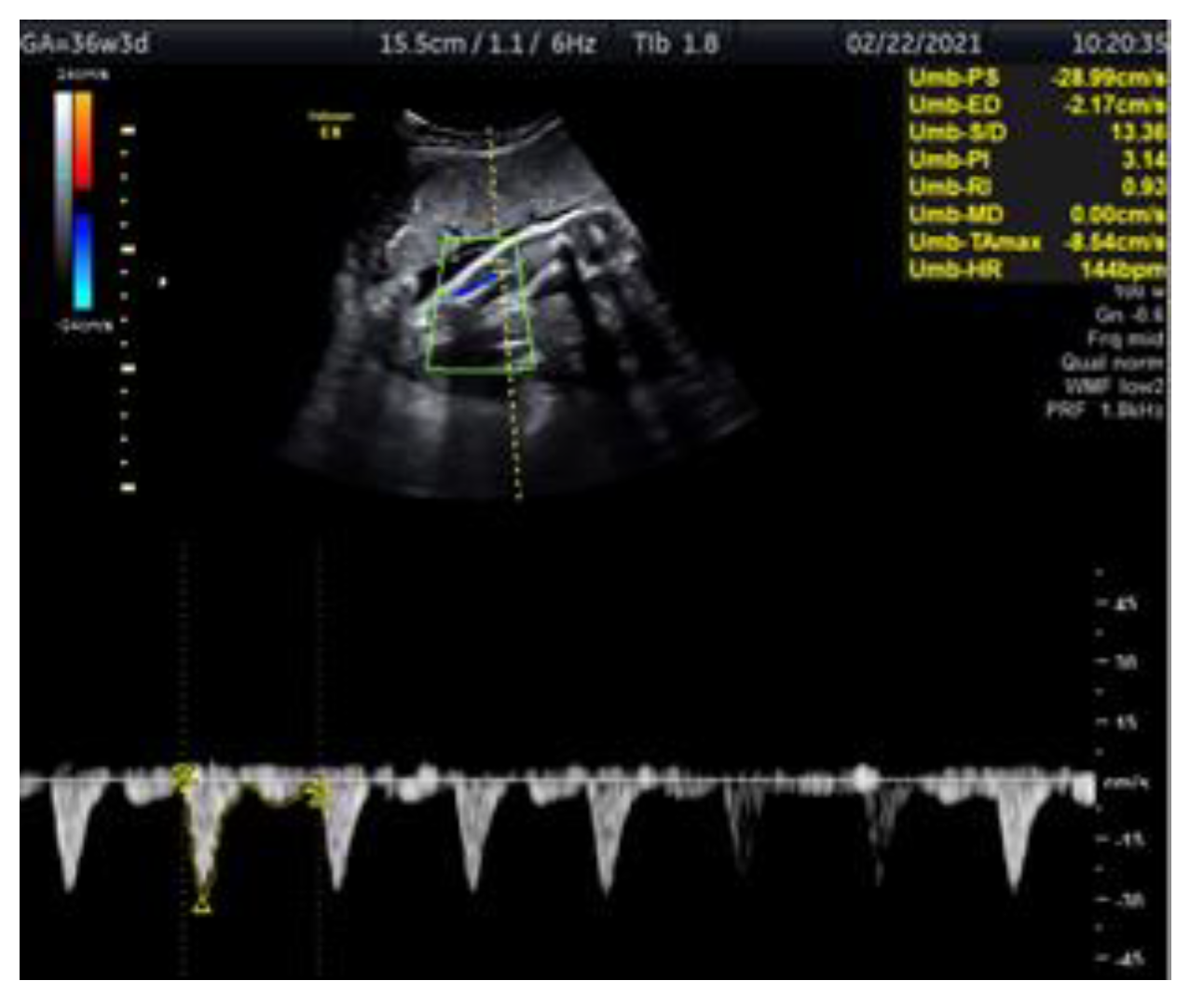
Medicina | Free Full-Text | Doppler Ultrasonography of the Fetal Tibial  Artery in High-Risk Pregnancy and Its Value in Predicting and Monitoring  Fetal Hypoxia in IUGR Fetuses
Medicina | Free Full-Text | Doppler Ultrasonography of the Fetal Tibial Artery in High-Risk Pregnancy and Its Value in Predicting and Monitoring Fetal Hypoxia in IUGR Fetuses
OB Images
OB Images

Male Fetus - 37+ Week || Ultrasound || Case 137 - YouTube

Resource Library search

Baby Within Calgary, 2D/3D/4D/HD Ultrasound Calgary

Doppler ultrasound in obstetrics - ScienceDirect

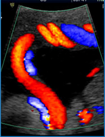
Normal pulsed and color Doppler of umbilical vein (37 WG) | Download  Scientific Diagram
Normal pulsed and color Doppler of umbilical vein (37 WG) | Download Scientific Diagram

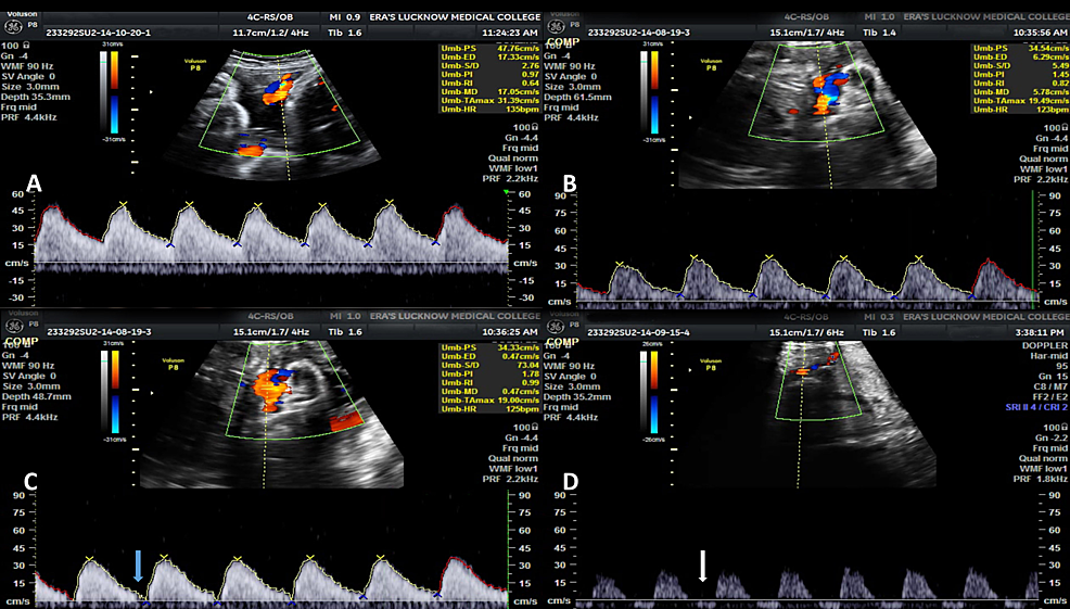
Cureus | Role of Color Doppler Flowmetry in Prediction of Intrauterine  Growth Retardation in High-Risk Pregnancy | Article
Cureus | Role of Color Doppler Flowmetry in Prediction of Intrauterine Growth Retardation in High-Risk Pregnancy | Article

Ophthalmic artery Doppler in prediction of pre‐eclampsia at 35–37 weeks'  gestation - Sarno - 2020 - Ultrasound in Obstetrics & Gynecology - Wiley  Online Library
Ophthalmic artery Doppler in prediction of pre‐eclampsia at 35–37 weeks' gestation - Sarno - 2020 - Ultrasound in Obstetrics & Gynecology - Wiley Online Library

Doppler ultrasound | FMF Certification | Welcome to the Fetal Medicine  Foundation
Doppler ultrasound | FMF Certification | Welcome to the Fetal Medicine Foundation

Pregnancy Week 37
Pregnancy Week 37

Doppler velocimetry of umbilical artery at 37weeks±4d | Download Scientific  Diagram
Doppler velocimetry of umbilical artery at 37weeks±4d | Download Scientific Diagram

37 Week Ultrasound! // How Big Is Baby? - YouTube

A Radiologist's Guide to the Performance and Interpretation of Obstetric  Doppler US | RadioGraphics
A Radiologist's Guide to the Performance and Interpretation of Obstetric Doppler US | RadioGraphics

Obstetric Ultrasound Normal Vs Abnormal Images | Fetal, Placenta, Umbilical  Cord Pathologies USG - YouTube
Obstetric Ultrasound Normal Vs Abnormal Images | Fetal, Placenta, Umbilical Cord Pathologies USG - YouTube

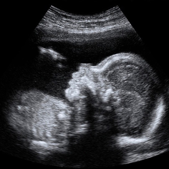
Fetal ultrasound images - 8 months - BabyCenter India

Umbilical Artery Doppler Assessment: A How-To Guide | Empowered Women's  Health
Umbilical Artery Doppler Assessment: A How-To Guide | Empowered Women's Health

Third Trimester Ultrasound Pictures

Ultrasound pregnancy Information | Mount Sinai - New York

Doppler ultrasound assessment of the ductus venosus in the normal fetus  between 20 and 40 weeks gestation in the Pakistani population - Syed Amir  Gilani, Amber Javaid, Alsafi Abdella Bala, 2010
Doppler ultrasound assessment of the ductus venosus in the normal fetus between 20 and 40 weeks gestation in the Pakistani population - Syed Amir Gilani, Amber Javaid, Alsafi Abdella Bala, 2010
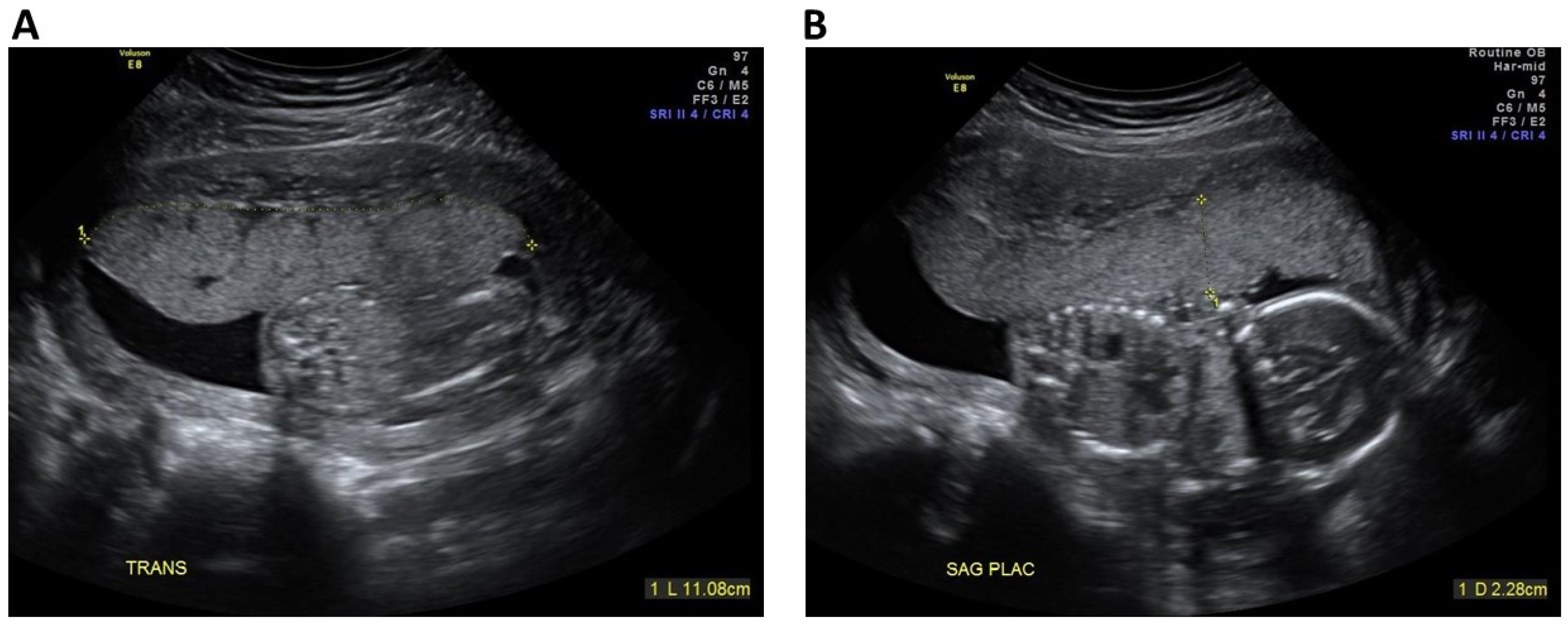
JCM | Free Full-Text | Contribution of Second Trimester Sonographic  Placental Morphology to Uterine Artery Doppler in the Prediction of  Placenta-Mediated Pregnancy Complications
JCM | Free Full-Text | Contribution of Second Trimester Sonographic Placental Morphology to Uterine Artery Doppler in the Prediction of Placenta-Mediated Pregnancy Complications

Wellbeing scan for babies - 32 weeks scan & 28 week scan. Doppler scan

First-Trimester Uterine Artery Doppler Analysis in the Prediction of Later  Pregnancy Complications
First-Trimester Uterine Artery Doppler Analysis in the Prediction of Later Pregnancy Complications

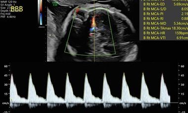
PDF] Doppler indices of the umbilical and fetal middle cerebral artery at  18-40 weeks of normal gestation: A pilot study. | Semantic Scholar
PDF] Doppler indices of the umbilical and fetal middle cerebral artery at 18-40 weeks of normal gestation: A pilot study. | Semantic Scholar